
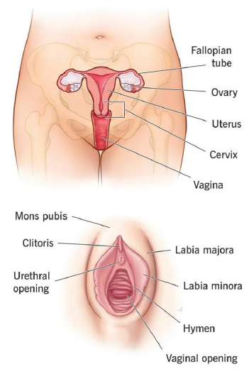
Again, this system consists of organs which allows for the production of
a new individual. The organs involved are the ovaries, fallopian tube,
uterus, cervix and vagina.
The Ovaries
The ovaries are the primary female reproductive organs, with each
one being a solid oval structure around the size and shape of an
almond.
They are approximately 3.5 cm in length and 2 cm wide and are
located one on each side of the uterus, in the lateral walls of the
pelvic cavity.
They are suspended by the ligaments in the upper pelvic cavity, one
being on either side of the uterus. At the birth of a female, each ovary
contains over 200,000 immature ova, with each ova being encased in
a sac called a follicle. In response to various sex hormones, each
follicle develops until they become primary follicles, which is when
ovulation occurs.
The ovum breaks free of the follicle and enters the fallopian tube. The
ovaries are also responsible for producing oestrogen and
progesterone, which are vital for proper reproductive function
There are two fallopian tubes, each about 4 inches in length and a
narrow as a piece of string, attached to a side of the uterus. Each
tube resembles a funnel, which is wider at the ovary and becomes
narrower at the uterus.
The fallopian tube has 20 to 25 fingers like projections (fimbriae) that
encourage the mature ovum to enter the fallopian tube. Once in the
tube, tiny hairs in the lining help push the ovum down the narrow
passageway towards the uterus.
If fertilization takes place, it will usually occur in the wider part of the
fallopian tube. Once the ovum has become fertilized it is called a
zygote and travels to the uterus over the next 7 days.
The Uterus
The uterus is suspended by broad ligaments and is situated between the
bladder and the rectum. It is shaped like an upside-down pear, with a thick lining, muscular walls and a rich blood supply. It is made up of three layers, being the peritoneum (outer layer), myometrium (middle layer) and endometrium (inner lining). The uterus serves as a pathway for sperm to reach the fallopian tubes as well as to be able to expand and contract to accommodate a growing foetus and push the baby out during labour. If fertilisation occurs, the uterus will provide a source of attachment and nourishment for the growing zygote, which embeds into the endometrium. When a woman isn’t pregnant, the uterus is only about 3 inches long and 2 inches wide and the lining of the uterus (endometrium) breaks down during menstruation.
The Cervix
The uterus ends at the cervix which is the lower portion or neck of the
uterus, which is lined with mucus and joins the top end of the vagina. It is a thick tube of smooth muscle that acts as a channel for sperm to reach the waiting ovum. During late pregnancy the walls of the narrowed channel thin out to allow for the babies head to descent. The opening of the cervix is very small and during childbirth, the cervix can expand to allow a baby to pass.
The Vagina
The vagina is the female’s sex organ and extends about 3 – 5 inches inside, up to the cervix. It is a muscular, ridged sheath connecting the external genitals to the uterus. The vagina acts as a pathway for the penis to enter during intercourse to allow sperm to be deposited. It also acts as a passageway for the birth of a baby. During sexual arousal, droplets of fluid appear along the vaginal walls and eventually cover the sides of the vagina completely. The tissues are rich in blood vessels which when engorged with blood as a result of sexual arousal, press against the tissue, forcing natural tissue fluids through the walls of the vagina.
Menstruation, Conception and Pregnancy
Menstruation is controlled by hormones and the cycle is usually a 28 day
process, although this can vary greatly.
- During day 1 – 5 the hormones oestrogen and progesterone reduce
which causes a breakdown of the endometrium. This results in the
discharge of blood, tissue fluid and mucus, but also in the
preparation of a mature follicle. - During days 6 – 13, two more hormones, Follicle Stimulating Hormone (FSH) and luteinizing hormone stimulate the ovaries to produce more oestrogen. This in turn allows for the endometrium to re-build.
- By now a mature follicle is ready to be released on day 14. Between days 15 – 28, progesterone is stimulated by luteinizing
hormone to prepare the uterus to receive the fertilised ovum, if there is one. - If fertilisation does not occur then the hormone changes will start the cycle over again and initiate the breakdown of the lining.
The process of conception is nothing short of a miracle. Once the ovum has been released, it travels along the fallopian tubes, producing an enzyme that attracts any sperm. One sperm will break through the tough coating of the ovum to fertilise it, which will then continue to the uterus. Once the zygote has arrived in the uterus it will implant itself into the endometrial lining and become an embryo.
Pregnancy
The first four weeks after fertilisation will show a rapid amount of
development as all of the major organs and body systems start to
develop. The placenta and umbilical cord, which sustains pregnancy,
are also being formed.
- During weeks 5 – 8, the embryo starts to become recognisably
human in form as the limbs begin to grow and the torso straightens
out. - By weeks 9 – 12, the first trimester is closing and the foetus is almost double in length with all organs formed. The eyelids are fused with the eyes remaining closed.
- Weeks 13 – 16 see the foetus rapidly increasing in size, with
movements alerting the pregnant women of its presence. The foetal
circulation is now established with the blood being pumped around
its body. - By the fifth month, the skin of the foetus has become more mature
and a network of blood capillaries and nerve endings has become
established. - The sixth month marks the end of the second trimester and the facial features are beginning to resemble those of a full term baby. The growth rate is slow but weight is being gained rapidly.
- The last trimester up until 40 weeks is a period where the foetus
continues to develop, but in a much smaller space. - By week 33, the foetus is still putting on weight at just over 5lb, but
still lacks adequate fat stores under the skin. - The last few weeks of pregnancy can be a time of great discomfort
for many women. For the foetus the fingernails have reached the
ends of the digits and its body is covered in a coating called vernix. - The average pregnancy lasts from 38 weeks from fertilisation or 40
weeks when calculated from the last day of the menstrual cycle.
