
The main function of the respiratory system is to allow oxygen to enter
the body and for carbon dioxide to leave. This is called “gas exchange”
and takes place on an internal level into tissues and an external level
into the lungs. It is vital that it takes place for life to continue.
The respiratory system not only deals with breathing and oxygenating
the blood, but also performs a wide range of additional tasks, including
cleans and filters the air that we breathe and making our voice more
resonant. In order for gas exchange to occur, the respiratory system’s
numerous organs must cooperate. The circulatory system collaborates
with this system to function. Through the mouth or nose, air is drawn
into the body and purified of undesired dust. Once in the trachea, it
passes via the rear of the pharynx and into the split bronchi, which lead
to the alveoli via the bronchioles. Gas exchange takes place just here in
the alveoli.
The respiratory system consists of:
- The nose
- The diaphragm
- The larynx
- The lungs
- The pharynx (throat)
- The trachea
- The bronchi
- The bronchioles
- The alveoli
The Mouth allows an intake of air if there is a high demand or if the
nasal passage is blocked in any way. It is an oval shaped cavity which is
lined with a mucous membrane. The mouth contains the soft and hard
palate, forming the roof of the mouth, as well as the gums in which the
teeth sit. It is not ideal to continually breathe through the mouth as the
air is not as well filtered and it can cause other medical problems.
The nasal cavity traps particles that enter the passages by containing
shelf-like structures called turbinate’s. Any material that is deposited in
the nose is transported by ciliary action to the back of the throat in
around 10-15 minutes. The vascular mucus membranes of the nose will
also warm and moisten the air as it is inhaled. The mucus which is
produced will also be moved to the back of the pharynx for either
swallowing or expectoration. The nose is formed by the two nasal bones
and by cartilage and is divided by a septum. The nose also acts as a
sounding chamber for the voice as some of the bones surrounding the
nasal cavity are hollow. These hollows are called paranasal sinuses and
allow the voice to become resonant, lighter and to secrete mucus to
help with air filtration.
The pharynx (throat) is a muscular cavity that begins from behind the
nose to the beginning of the voice box and the oesophagus. The pharynx
is divided into three sections. The nasopharynx lies behind the nose and
can be seen when the mouth is wide open, the oropharynx which lies
behind the mouth, and the laryngopharynx which lies behind the larynx.
The upper part of the pharynx lets air pass through, whilst the lower
parts permit air, foods and fluids to pass. When it is necessary to
swallow, breathing will stop as the oropharynx becomes blocked off
from the nasopharynx as the soft palate is raised, as it is impossible to
be able to breathe whilst swallowing.
The larynx, also known as the voice box, is a 2” tube shaped structure
which is located at the entrance of the trachea. The larynx contains two
vocal cords, which will vibrate together when air passes between them.
This gives us the sound of the voice. The larynx is made up of several
irregular cartilages and the lobes of the thyroid gland are on either side.
The oesophagus, which is the tube that carries food from the mouth to
the stomach, is just behind the trachea and the larynx. Both openings of
the oesophagus and the larynx are close together in the throat, so when
the act of swallowing occurs, a flap called the epiglottis keeps the food
out of the windpipe by moving down over the larynx.
The trachea, also known as the windpipe, is a tube like structure
consisting of between 16 – 20 rings of cartilage that joins the nose and
mouth to the lungs. It measures approximately 10-12” in length and runs
from the lower part of the larynx to the lungs by dividing into the
bronchi. The trachea contains an epithelial lining that secretes mucus,
which traps any dust. It is then swept upwards by the cilia towards the
larynx away from the lungs.
The bronchi are supported by cartilage and are formed when the
trachea forks into two branches, making up the left and right bronchi.
These branches then divide again, with the right Bronchus being wider
and shorter than the left. The right bronchi then divide into two
branches for the middle and lower lobes. The left bronchi is nearly
double in length, being 5cm long and divides again, one for each
broncho-pulmonary segment. Within the lungs, the bronchi divide again
into smaller bronchi, called bronchioles. There are numerous glands in
the wall of the bronchi which secrete slimy mucus, which helps to trap
dust and any other particles, which are then propelled upwards to the
mouth by cilia.
The bronchioles are the first divisions of the bronchi that no longer
contain cartilage, but are made up of a single layer of epithelial cells.
The bronchioles are smaller than one millimetre in diameter and control
the air distribution into the lungs. The bronchiole end in the alveoli.
The alveolar sac contains around 300 million alveoli, which are arranged
in grape like clusters to increase the surface area, which can become
reduced due to irritants such as dust. It is here that gas exchange takes
place. To allow this to happen, the alveoli are constantly moist and are
surrounded by a network of capillaries. Oxygen is in a higher
concentration in the alveoli than in the blood and so therefore it is able
to diffuse into the blood through a thin layer of cells. The reverse
happens with carbon dioxide, which is a higher concentration in the
blood than the alveoli and so it diffuses into the alveoli through the thin
layer of cells.
The lungs are located in the thorax and are cone shaped. They make up
one of the largest organs of the body with a huge surface area. The
main role of the lungs is to exchange gas; oxygen for carbon dioxide and
on average a person breathes 25,000 times a day, moving 10,000 litres of
air a day.
Mucus is produced in the lungs that traps any inhaled particles, which
can be removed by coughing.
The lungs are situated in a space, known as the pleural cavity. Each
lung is covered in two thin layers of a single celled membrane called
pleura which slide back and forth over each other every time a breath is
taken to allow the lungs to expand and contract. There is a small
amount of fluid here to prevent friction. The pleura, which are connected
to the chest wall are called the parietal pleura, and the pleura that are
attached to the lung are called visceral pleura.
The front and back of the lungs are protected by the ribs, and the
intercostals muscles help allow the chest wall to move. The front of the
ribs contains costal cartilage which connects the sternum and the ends
of the ribs. The back of the lungs contains the transverse processes of
the thoracic vertebrae. The lungs differ on either side with the right lung
having 3 lobes; the superior, middle and inferior lobe and the left lung
only having the superior and inferior lobe.
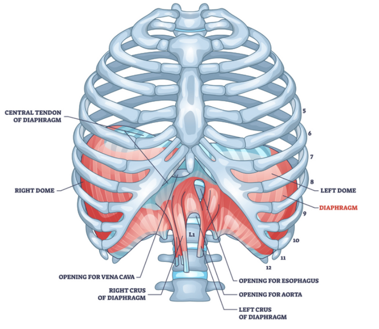
The Diaphragm is a dome shaped muscular sheet that extends along
the bottom of the rib cage and inserts into the lower ribs. The
diaphragm relaxes during inhalation to allow more room in the thoracic
cavity, which in turn creates a suction to allow air to be drawn into the
lungs. When you exhale, the diaphragm expands which reduces the
amount of space in the cavity for the lungs, which forces the air out.
The Intercostal Muscles occupy the space in-between the ribs and are
made up of two types. The internal muscles are on the inside of the rib
cage and extend from the front of the ribs and go around the back, and
the external muscles are on the outside of the ribs and cover the back of
the rib, around to the bony part at the front. They receive messages from
the brain to control breathing, and are responsible for working
alongside the diaphragm.
Breathing Mechanism
To be able to take in oxygen and allow carbon dioxide to be expelled, a
complex procedure needs to take place.
Inhalation:
- The diaphragm contracts and moves downwards This forces the rib
cage muscles to contract - The ribs then move up and out
- There is decreased pressure in the chest
- The air is sucked down into the lungs through:
- Nose, pharynx, larynx, trachea, bronchus, bronchiole and to the
alveoli - Once the oxygen is in the alveoli, gas exchange takes place so that
the carbon dioxide is ready to be exhaled. The reverse then happens.
Exhalation:
- The muscles of the diagram and intercostals relax The size of the
thorax reduces - Air is forced out of the lungs
Gas Exchange
Once the air that we have inhaled reaches the lungs, the 21% of
dissolved oxygen then diffuses through the alveolar lining cells of the
alveolar and the walls of the capillaries and enters the plasma of the
blood.
From the plasma, the oxygen then diffuses into the red blood cells
(erythrocytes) and combines with the haemoglobin to form
oxyhaemoglobin.
The newly oxygenated blood then leaves the capillary network and
enters the pulmonary veins to be transported back to the heart to be
pumped around the body for its use.
Once the oxygen has travelled the body, the deoxygenated blood leaves
via the capillary network from the pulmonary artery back into the
alveoli.
The exhaled breath still contains 16% oxygen and 4
