
The main functions of this system are to supply oxygenated blood
throughout the body, and to remove waste products, such as carbon
dioxide. It is able to carry out this task by using three organs; the blood,
vessels and the heart.
Blood – If we think of the circulatory system as a transportation service
then the blood would be the bus. Carrying and distributing oxygen,
nutrients, antibodies, heat and hormones, it travels through the body,
whilst also collecting waste products, such as carbon dioxide, which
need to be removed. The blood is made up of 4 components and an
adult has 10.6 pints.
Plasma is a straw coloured fluid and accounts for about half of the total
volume of blood. It is necessary for the suspension of blood cells and is
made up of 90% water. The major protein in plasma is albumin which
prevents fluid from leaking out of the blood vessels into tissues. Plasma
also supplies water when additional liquids are needed in the tissues of
the body, as well as play a crucial role in regulating the body
temperature by carrying heat around the body. The Plasma contains
dissolved substances, most of these are useful and are carried to places
where they are to be stored or used. The products of digestion including
glucose, amino acids, mineral salts and vitamins are carried from the
small intestines (ileum) to other organs. Without plasma, the life-giving
blood cells would be left without transportation.
Red blood cells (erythrocytes) carry oxygen, which is needed by the cells
to produce energy, and are formed in the bone marrow of long bones.
They are the most common type of blood cell and live for around 12o
days and make up around 40% of the bloods volume. These blood cells
contain a protein chemical called haemoglobin which is bright red in
colour. Haemoglobin allows the oxygen to be collected in the lungs by
binding its molecules with the oxygen and then distributes it around the
body. Carbon dioxide is then collected to allow it to be removed. If you
have a lack of haemoglobin, you may develop a condition called
anaemia.
White blood cells (leucocytes) are involved in the protection of the body
and are on the continual look out for any sign of bacteria. There are five
main types of white blood cells which all have a differing role. The white
blood cells that are most numerous are Neutrophils which kill and
ingest foreign material. Lymphocytes help protect against viral
infections and produce antibodies. Monocytes ingest dead and
damaged cells, Eosinophils protect by killing parasites and destroying
some cancer cells as well as being involved in the allergic response, as
well as the basophils.
White blood cells have a shorter life expectancy than red, only surviving
for about 3 weeks. A drop of blood can contain anywhere from 7,000 to
25,000 white blood cells at a time. If an invading infection fights back
and persists, that number will significantly increase.
Platelets, also called thrombocytes are necessary for the blood clotting
process to take place. They are irregularly-shaped and colourless and
have a sticky surface that lets them form clots to stop bleeding. When
you cut yourself, platelets in the blood react to the air and calcium,
vitamin K, and a protein called fibrinogen are released. This forms a
blood clot, which seals or plug’s the hole and later on becomes a scab. A
scab is an external blood clot that we can easily see, but there are also
internal blood clots. A bruise, or black-and-blue mark, is the result of a
blood clot. Clotting is necessary, but sometimes it can be very dangerous
as if a blood clot forms inside of a blood vessel, it can block the flow of
blood, cutting off the supply of oxygen.
Blood Vessels
If the blood acts as a bus, then the blood vessels are road networks that
it travels along. There are three main vessels, and the blood follows two pathways known as pulmonary and systemic.
Arteries always carry blood away from the heart, with the exception of
the pulmonary artery (we will look at that later). They are the biggest of
the vessels and carry oxygenated blood. The walls of the artery are
muscular and elastic which helps allow the blood to travel the body. The
aorta is the largest artery of the body, which originates from the heart,
and branches out into smaller arteries, called arterioles which branch
into capillaries. An artery has three layers. An outer layers of tissue a
muscular middle and an inner layer of epithelial cells. There are two
types of arteries. Pulmonary arteries carry blood from the heart to the
lungs and systemic arteries carry blood to the rest of the body. The
smallest arteries are called arterioles and deal with delivering blood
from the arteries to the capillaries
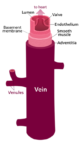
Veins carry deoxygenated blood to the heart, under low pressure, in
order for it to get sent to the lungs. Veins contain valves, which act like
doors – preventing the blood from flowing in the wrong direction. The
largest vein is the vena cava which leads to the right atrium of the
heart. Veins also have three layers: an outer layer of tissue, muscle in the
middle, and a smooth inner layer of epithelial cells, but the layers are
thinner and contain less tissue. Because it lacks oxygen, the blood that
flows through the veins has a deep red colour. The walls of the veins are
rather thin which makes the blood visible through the skin on some
parts of the body, such as the hands, wrists and ankles. As the skin
refracts light, the deep red colour actually appears a little blue from
outside the skin. Veins can be classified into four different types.
Pulmonary veins carry blood from the lungs to the left atrium of the
heart. Systemic veins carry deoxygenated blood from the remainder of
the body to the right atrium of the heart. Superficial veins are to be
found close to the surface of the skin and deep veins are located deep
within muscle tissues.
Capillaries are very small vessels that transport blood from the arteries
to the veins. They have thin walls, made up of endothelium (single layer
of overlapping flat cells) that allows substances such as nutrients to
exchange. The capillaries are so small that red blood cells have to travel
through them in single file. The flow of blood through the capillaries is
controlled by structures called precapillary sphincters, which are located
between arterioles and capillaries. They contain muscle fibres that allow
them to contract. Blood flows freely to the capillary beds of body tissue
when the sphincters are open, but when the sphincters are closed blood
is not allowed to flow. Plasma moves out of the capillaries and becomes
tissue fluid. This fluid bathes the cells in nutrients and oxygen, some
waste and excess fluids move into the lymphatic vessels, with the
carbon dioxide and waste returning to the capillaries.
Veins carry deoxygenated blood to the heart, under low pressure, in
order for it to get sent to the lungs. Veins contain valves, which act like
doors – preventing the blood from flowing in the wrong direction. The
largest vein is the vena cava which leads to the right atrium of the
heart. Veins also have three layers: an outer layer of tissue, muscle in the
middle, and a smooth inner layer of epithelial cells, but the layers are
thinner and contain less tissue. Because it lacks oxygen, the blood that
flows through the veins has a deep red colour. The walls of the veins are
rather thin which makes the blood visible through the skin on some
parts of the body, such as the hands, wrists and ankles. As the skin
refracts light, the deep red colour actually appears a little blue from
outside the skin. Veins can be classified into four different types.
Pulmonary veins carry blood from the lungs to the left atrium of the
heart. Systemic veins carry deoxygenated blood from the remainder of
the body to the right atrium of the heart. Superficial veins are to be
found close to the surface of the skin and deep veins are located deep
within muscle tissues.
